General Muscle Strains
Overview
Muscle strains, also known as pulled muscle, usually arise from an indirect insult from application of excessive tensile forces. Most muscle strain injuries occur from a powerful eccentric contraction or overstretching of the muscle, while more severe injuries may involve partial or complete tears in tissues. Muscle strains are one of the most common injuries, particularly in sport where 90% of all sports-related skeletal muscle injuries account as muscle strains. For most with grade I muscle strains, healing takes about 2-4 weeks, and typically 2 months for those with a grade II strain. In rare and severe instances, grade III strains could take at least 6 months, or longer, depending on the type of surgery received. Muscle strains are predisposed by older age, previous muscle injury, less flexibility, lack of strength, and fatigue. Minor muscle strains typically heal on their own with rest, however therapeutic massages could speed a strained muscle injury, by helping to loosen the tight muscle and increase blood flow to help heal damaged tissues.
Anatomy
These kind of injuries mostly occurs at the musculotendinous junction (primary site of force transmission between the two tissues), where the tendon emerges from the muscle belly and myo-tendinous junction. During eccentric muscle actions, or when muscle tension increases suddenly, the damage may occur in the area beneath the epimysium and the site of muscle attachment to the periosteum. The region adjacent to the MTJ is more susceptible to injury than any other component of the muscle unit, respectively, from type and direction of applied forces and muscle architecture. Haemorrhage occurs in the affected area, up to 24 hours after injury, with an inflammatory reaction occurring after. Laying down of fibrous tissue and scar tissue starts after 7 days, being visible after 2 weeks.
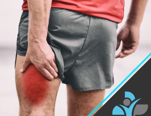
Symptoms of the pathology
• Pain, tenderness, redness, or bruising
• Limited range of motion
• Muscle spasms
• Swelling
• Localised pain and general muscle weakness
The inflammatory phase- occurs within a few hours however peaks 1 to 3 days after the injury. Redness, swelling, heat, pain, decreased range of motion.
The Proliferation phase- 24 to 48 hours after the injury. If a muscle is partially torn, this phase will repair the tear by laying down new fibres to repair that gap (scar tissue).
The Remodelling Phase- up to 1-2 years. Formation of the muscle where re-injury is more susceptible.
Causes
Muscle strains usually occur from an indirect insult, from application of excessive tensile forces. The most commonly injured muscles are the bicep femoris, rectus femoris, and the medial head of the gastrocnemius, all with a greater percentage of type II fibres, a pennate architecture, cross two joints. Strains typically occur during the eccentric phase of a muscle action or during excessive loading, where the muscle can become overstretched beyond its limit. Laboratory studies show that partial and complete injuries exhibit disruption of muscle fibres near the muscle-tendon junction, where tissues tear when forces across the musculotendinous unit contract too strongly.
Risk factors: Muscle imbalances, poor conditioning (e.g., weaker muscles), fatigue in the muscles.
Diagnosis
Manual Testing – observation, palpation, strength testing, and evaluation of motion.
Most muscle strains can be diagnosed through manual testing, where pain is typically felt by the patient with resisted muscle activation, passive stretching, and direct palpation over the muscle strain. Assessing tenderness, any palpable defect, and strength at the onset of muscle injury will determine grading of the injury and provide direction for further diagnostic testing and treatment.
Often, diagnosis is uncertain and further detail is needed to locate the muscle strain. Radiographs, ultrasound (US), and magnetic resonance imagine (MRI) are common imaging tools. Radiographs would return normal in acute muscle strains, however, may be useful in differentiating between bony and muscular aetiologies of pain.
Clinical grading system
Grade I- localised pain worsening with movement, mild swelling, tenderness, and minimal haemorrhage. (< 10° RoM deficit) Grade II- localised pain worsening with movement, substantial pain to palpation, considerable pain on contraction with greatly disturbed gait. (10-25° RoM deficit) Grade III- (muscle or tendon rupture) severe pain, swelling, and haematoma present. Palpable defect and loss of muscle function. (> 25° RoM deficit).
Treatment
Treatment for muscle strain injuries has remained the same over the years, with little scientific basis for most treatment protocols. Instead, it provides a basis for the currently accepted methods of treatment.
Initial treatment consists of rest, ice, compression, and nonsteroidal anti-inflammatory drug therapy. As pain and swelling subside, physical therapy should be initiated to restore flexibility and strength. Strengthening, range of motion, proprioceptive exercises, and functional training are subsequently followed, that should progress gradually. Stretching exercises should be done carefully without pain, and only to the point of discomfort. Strengthening exercises should progress sequentially through isometric, isotonic, isokinetic, and functional exercises, through a pain free range of motion. Massage therapy may also help to relax injured muscles and improve range of motion, and immobilisation therapy can be used to remain the injured area in a neutral position.
Exercises
The type and intensity of exercises will depend on the injured area and should be performed through a pain-free range of motion and only to a point of discomfort.
For the most common muscle strain injuries, examples include:
Hamstrings (add resistance in absence of pain):
– Hamstring curl- Lie on stomach, lift foot of affected leg by bending the knee
– Hip extension- Face a wall with hands at about chest level. Kick the affected leg behind you, remaining in control
Quadriceps (can add ankle weight to increase difficulty)
– Straight leg raise (laterally rotated)- raise leg parallel to the bent leg without arching the back.
– Wall squat- slowly lower body down and hold, maintaining pelvis, back, and head against the wall.
Gastrocnemius
– Plantar flexion with resistance- point the foot away while sitting down, holding a loop of resistance band to apply resistance
– Calf raises- seated in the early stages or standing in later stages. Raise up onto toes and lower the heels back down.
If you are suffering from any of the things listed above, you can contact us through an email info@livewellhealth.co.uk or give us a call on 0330 043 2501.